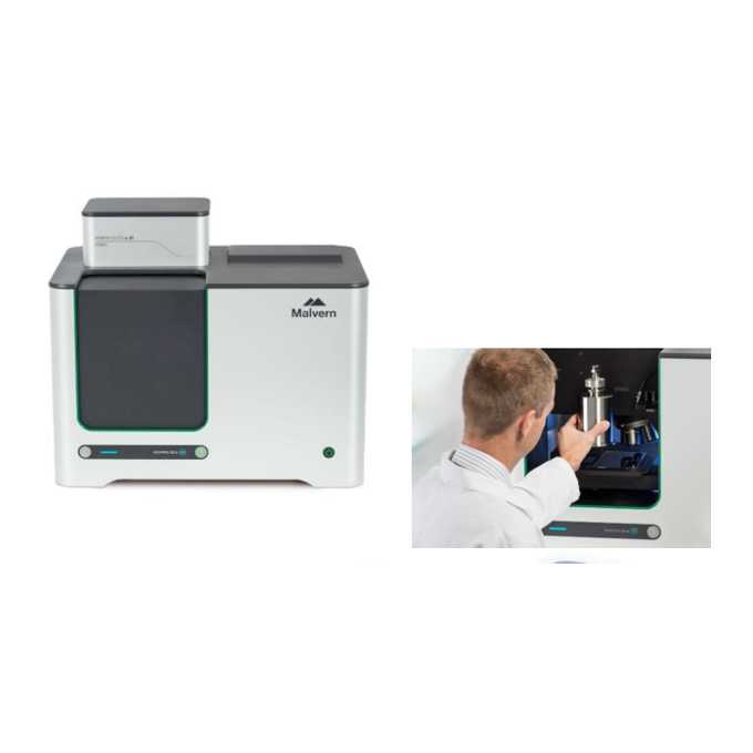
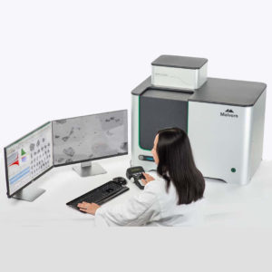
Morphological imaging is fast becoming an essential technology in the laboratory toolkit for particle characterization. Providing automated, rapid, and component-specific morphological information, Morphologi instruments are employed to solve formulation and deformulation challenges, optimize material properties, and create confidence throughout development and manufacturing. These systems enable process control and optimization and provide quick identification of the cause of process deviations. Morphological imaging applies the technique of automated static image analysis to provide a complete, detailed description of the morphological properties of particulate materials. By combining particle size measurements such as length and width, with particle shape assessments such as circularity and convexity, morphological imaging fully characterizes both spherical and irregularly-shaped particles. This enables deeper understanding of a sample’s characteristics through precise detection of agglomerates, foreign particles and other anomalous materials. It also delivers the data required to cross-validate other particle sizing methods which apply an equivalent-sphere approach to reporting particle size distributions.
Features
Broad particle size range from 0.5 μm to >1300 μm enables size measurements of a wide range of samples
20+ morphological parameters deliver a highly-detailed description for deeper understanding of your particulate material
SOP control, from sample dispersion to data analysis, provides simple and automated operation for robust, repeatable measurements
Automated ‘Sharp Edge’ analysis enables detection of even low contrast particles
High resolution microscope ensures quality particle images for optimum image analysis data
Integrated dry powder dispersion unit delivers reproducible sample dispersion, critical to achieving meaningful results
Dedicated sample presentation accessories enable measurement of a wide variety of sample types, including suspensions and filters
Advanced data exploration tools generate maximum sample knowledge
Advanced manual microscope mode and ability to return to particles of interest enables an even closer examination of unexpected particles
21 CFR Part 11 software option ensures regulatory compliance
Specifications
Technology Morphologi 4 : Static automated imaging
Morphologi 4-ID : Static automated imaging combined with Raman spectroscopy
Morphological analysis Static automated imaging
Particle size range 0.5 μm – 1300 μm (upper limit may be extended for some applications*)
Particle properties measured Size, shape, transparency, count, location
Particle size parameters Circle equivalent (CE) diameter, length, width, perimeter, area, maximum distance, sphere equivalent (SE) volume, fiber total length, fiber width
Particle shape parameters Aspect ratio, circularity, convexity, elongation, high sensitivity (HS) circularity, solidity, fiber elongation, fiber straightness
Particle transparency parameters Intensity mean, intensity standard deviation
Integrated sample dispersion unit for fully automated dispersion and measurement of dry powders. Manual or SOP control of dispersion pressure, injection time and settling time
Illumination White light LED: brightfield, diascopic and episcopic; darkfield, episcopic
Detector 18 MP; 4912 x 3684 pixel color CMOS array; pixel size 1.25 μm x 1.25 μm
Optical system Nikon CFI 60 brightfield / darkfield system
Lens 2.5x 5x 10x 20x 50x
Particle size range in μm by lens (nominal): 8.5-1300 4.5-520 2.5-260 1.5-130 0.5-50
Software
Build-it data evaluation.
Intelligent neural network system also suggests how to improve results
Built-in tools for method development according to ISO-13320.
Adaptive Correlation to determine optimum measurement duration
21 CFR Part 11 Enables an operating mode that assists with ER/ES compliance.
Dimensions
Dimensions 810 mm (W) x 520 mm (D) x 685 mm (H)
Weight 80 kg
Power requirements 100-240 V ac 50/60 Hz (<100W load)


